Groin Pain: Why the Hip Joint Hurts in the Groin, Not the Hip
Published: Author: Mikheil Mikeladze, MD
Groin pain is the most typical sign of a hip joint problem, because the joint itself sits deep in the groin. What everyday speech calls “the hip”, meaning the bony bump on the outer side, is several centimetres to the side of the joint and above it. It is this anatomical confusion that leads a patient to a doctor complaining about a “knee” or a “back” while the problem is in the hip joint.
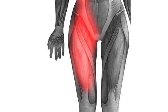
Where the hip joint actually sits
The hip joint lies deep inside the body, below and slightly to the side of the groin crease, under a thick layer of muscle. You cannot feel it with your hand: unlike the knee, which sits just under the skin, it cannot be palpated with the fingers.
On the outer side of the hip you can clearly feel a bony prominence, the greater trochanter. This is the part of the femur where the tendons of the gluteal muscles attach. The greater trochanter is not part of the joint, and putting a hand on it tells you nothing about the joint.
The distance between the two points is small, but anatomically they are different worlds. The joint is inward and forward, the greater trochanter outward and back. Between them lie several layers of muscle, tendons and bursae, and each of these has its own pattern of pain.
This difference is where the common mistake comes from. When someone says “my hip hurts” and puts a finger on the bone at the side, they are usually not talking about the joint. The reverse happens too: people with groin pain often assume it is a hernia or a pulled muscle and never think about the joint at all.
That is why in consultation I ask the patient to put a hand on the place where it hurts most. That simple movement often says more than a long description.
Why joint pain is felt in the groin
Joint pain is felt in the groin because the joint capsule and its sensitive nerve endings are concentrated on the front surface of the joint, and that surface faces the groin.
The pain rarely stays in one spot. It usually spreads across the front or inner surface of the thigh and sometimes reaches the knee. Many patients describe it with a sweeping gesture: they run a hand from the groin down the thigh and say that it “goes this way”.
The character of the pain is mechanical. Walking, standing for a long time and climbing stairs make it worse; rest eases it. Deep flexion of the joint, such as sitting down in a low chair or tying shoelaces, often makes it sharply worse.
Groin pain has other causes too, and they have to be considered. An inguinal hernia, irritation where the abdominal muscles attach, a tendon problem after sport, and in women the state of the pelvic organs all produce complaints in the same area. What sets joint pain apart is that it is tied to movement and usually eases at rest.
The other early signs of hip osteoarthritis are set out in detail in a text of their own: the early signs of hip osteoarthritis that people overlook.
Why the knee hurts when the problem is in the joint
Pain referred to the knee is the result of a signal from the hip joint: both areas are served by shared nerve pathways and the brain cannot always locate the source precisely. This phenomenon is called referred pain.
The practical consequence is simple. A patient goes to a doctor with knee pain, the knee is imaged, and the image shows nothing significant. Treatment stays aimed at the knee and months pass without result. I hear this story often in my consultations.
There is a simple guide: if the knee looks normal, is not swollen, does not lock and is not tender on examination, yet the pain is still there, it makes sense to check the hip joint. Particularly if there is stiffness in the groin at the same time.
Hip osteoarthritis presenting as isolated knee pain is not rare, and I meet it regularly in consultation.
Children and adolescents need particular attention. Knee pain that appears at this age together with a limp always calls for examination of the hip joint, because several conditions of this period begin exactly like this and a timely diagnosis is critical in them.
What pain on the outer side of the hip means
Pain on the outer side of the hip, around the greater trochanter, is usually linked to irritation of a tendon or a bursa, the cushion that sits between tendon and bone, rather than to the joint cartilage itself.
This pain has its own signs:
- it can be felt with a finger: pressing on the bone is painful;
- it is worse when lying on that side and often wakes people at night;
- it appears when getting out of a car sideways or after standing for a long time;
- the range of motion in the joint is usually preserved.
There is still a link between the two conditions. A gait altered by joint arthritis loads the gluteal muscles unevenly, and that irritates the tendon. So a patient often has both pains at once, and treating only one does not give full relief.
The practical conclusion is that pain on the outer side does not cancel the need to check the joint. In consultation I check both directions, because the treatment plan depends on which source is the main one and which is secondary.
The full picture of hip joint disease, with causes and stages, is set out on the hip arthritis page.
What pain in the buttock and lower back means
Pain sitting in the back of the buttock and in the lower back more often comes from the lumbar spine, although the hip joint can present this way too. The two are told apart clinically.
The typical picture of pain coming from the lower back is this: it runs from the buttock down the back of the thigh, continues below the knee and comes with numbness, burning or weakness. Joint pain, by contrast, sits further forward and rarely goes past the knee.
There is a situation where both sources are present at once and mask each other. In that case the examination proceeds step by step: first we establish which movement provokes the pain, then imaging is added as needed.
| Site of the pain | Most likely source | Additional signs |
|---|---|---|
| Groin, front of the thigh | The hip joint | Restricted movement, morning stiffness |
| Front of the knee, with no other complaint | Referred pain from the joint | The knee looks entirely normal |
| The bony bump on the outer side of the hip | Tendon or bursa | Tender to touch, hard to lie on that side |
| Buttock, back of the thigh | The lumbar region | Numbness, burning, pain below the knee |
| Deep, one-sided pain in the buttock | Sacroiliac joint or the back of the hip joint | Worse after sitting for a long time |
The table is a guide and does not replace a diagnosis. The same person can have several sources at once.
How we try to tell the source apart
Telling the source apart starts with examination, not with imaging. In consultation I first check the movements that load the joint directly.
The most informative is internal rotation of the hip: with the knee bent, taking the shin outwards rotates the hip inwards. Restriction of this movement and pain during it point to the joint. If the same movement is free and painless, hip osteoarthritis is less likely.
Three simple questions are useful for observing at home:
- Is it hard to put on a sock or tie a shoelace on that side? This points to restriction in the joint.
- Is pressing on the bone at the outer side of the hip painful? This strengthens the tendon or bursa direction.
- Does the pain run below the knee, together with numbness? This justifies thinking about the lower back.
None of the three questions makes a diagnosis. Their purpose is to help you bring a more precise description to the consultation.
Keeping a record over a week is also useful: when the pain appears, what makes it worse and what helps. Such a record often explains more in consultation than a description of one day’s sensations, because the mechanical rhythm typical of osteoarthritis only becomes visible across several days.
Which investigation answers this question
The answer usually comes from a pelvic X-ray taken standing, under load. The image shows the width of the joint space, changes in bone density and osteophytes, the bony outgrowths that form on the surface of the bone.
One detail matters: the image should cover both joints and not only the painful one. Comparison with the other side often makes a change obvious that could go unnoticed on an image taken in isolation.
Further imaging is ordered when the X-ray does not explain the complaint. Magnetic resonance imaging shows the soft tissues, the blood supply to the femoral head and the surroundings of the joint, none of which appear on an X-ray. This is particularly necessary when the pain is severe and the image is quiet.
There is one more, highly informative method. If a local anaesthetic injected into the joint temporarily reduces the pain sharply, that directly confirms the source of the pain is the joint itself. Such a diagnostic block is used when the complaint and the image do not match and an answer is still needed.
At BAU International Hospital in Batumi I look at the image together with the patient and explain what I see. The full range of treatment, from conservative to surgical, is described on the treatment page.
What to remember
- The hip joint sits deep in the groin, not on the bony bump at the side of the hip.
- Groin pain running down the front of the thigh is the most typical complaint from the joint.
- Isolated knee pain sometimes comes from the hip joint.
- Pain on the outer side of the hip is more often a tendon or bursa matter.
- The site of the pain gives a direction; examination and an X-ray give the diagnosis.
Groin pain you have had for months without an answer deserves to be sorted out properly. At a consultation we will work out together where it comes from: book a consultation.
Common questions
If my hip hurts at the side, does that mean the joint is healthy?
No, that conclusion does not follow. Pain on the outer side of the hip is more often linked to an irritated tendon or bursa, but that problem is frequently the result of a gait already altered by the joint. Both can be present at once, so the examination looks in both directions.
Can hip osteoarthritis show up only as knee pain?
Yes, that happens, and it is not rare. The hip joint and the front of the knee share nerve pathways, so the brain reads a signal from the joint as knee pain. In these cases examining the knee and imaging it usually show nothing.
How do I tell whether the pain comes from the joint or the lower back?
Pain coming from the lower back usually runs down the back of the buttock, continues below the knee and comes with numbness or a burning feeling. Joint pain sits further forward, in the groin, and is made worse by bending or rotating the leg. The final answer comes from examination.
Do I still need an X-ray if the pain is clearly in the groin?
An X-ray is needed, because the site of the pain only points the doctor in a direction. The image shows the state of the joint space, whether there are changes in the bone, and whether this is a different condition altogether, one with a different timescale and different treatment.
This text is educational and does not replace a consultation. A diagnosis and a treatment plan can only be set after a personal examination.
Have a question about your own case?
At the consultation we look at your X-ray together and I tell you what stage the joint is at and what the next step is. Seeing a surgeon does not mean surgery.
More in short videos: follow me
Read next
- Hip arthritisOsteoarthritis or Arthritis: The Difference Between the Two DiagnosesOsteoarthritis is mechanical wear of the joint cartilage, while inflammatory arthritis is inflammation of the joint. The difference decides who treats you.
- Hip arthritisThe Early Signs of Hip Osteoarthritis That People OverlookThe first signs of hip osteoarthritis are rarely severe pain. Stiffness in the groin and trouble putting on socks usually appear months or years earlier.
- Non-surgical treatmentSeeing an orthopaedic trauma surgeon: which symptoms warrant a visitAn orthopaedic trauma surgeon treats joint, bone and muscle problems. Which symptoms warrant a visit, what a consultation covers and when not to wait.
